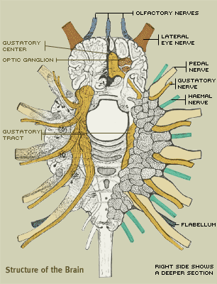
Structure of the Brain
At the top of the brain are three olfactory nerves (shown in blue) that lead to the horseshoe's olfactory sense organ. Immediately following these are the lateral eye nerves that connect to the crab's two lateral eyes.
The five pairs of pedal nerves (highlighted in tan) are large and complex bundles of fiber. One of the most striking features of the brain are the large nerve roots (shown in yellow). These are the gustatory nerves and supply the taste organs in the coxal spurs, located on the appendages. The gustatory organs are widely distributed over the neural surface of the head, but they are most widely developed in the appendages that come in frequent contact with the food.
The sixth nerve of this type (shown in blue) leads to the flabellum, which is a large spatulate organ that forms part of the crab's "pusher leg" (See Appendages for more). The function of the flabellum is to test the composition of the water passing to the gill chamber.
The haemal nerves, in green, contain motor and sensory fibers and are distributed mainly to the membrane and other tissues.

Illustration (modified) by William Patten